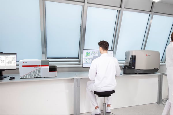
DxFLEX Flow Cytometer
The DxFLEX Flow Cytometer streamlines high-complexity testing with a 13-color* capability and new detector technology for easier compensation. Our innovative APD (avalanche photodiode) detectors offer advanced sensitivity and improve your diagnostic accuracy and keep your lab at the forefront of clinical flow cytometry.
High-Complexity Testing Made Easy
- Simplified compensation procedure (gain-independent compensation)
- More information per tube with up to 13 colors*
Advanced Sensitivity and Resolution
- Separation of dim populations
- Easier and robust gating
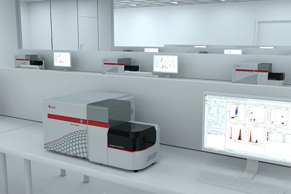
Compact Design
- Saves lab space
- No compressor cart
*In the US, the DxFLEX flow cytometer is cleared for 10-color in vitro diagnostic use with the ClearLLab 10C Reagent System. Fluorescence channels FL11-FL13 and all other applications are for Research Use Only.


DxFLEX Flow Cytometer Features
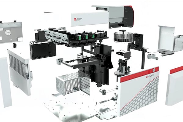
A compact design, intuitive software, up to 13-color detection and an enhanced optical path for increased sensitivity ensure accurate results and makes flow cytometry data collection easy even for novice users.
Simplified compensation
The gains of the APD detectors used in the DxFLEX flow cytometer can be calibrated for a linear response – leading to the predictability of the signals.
- A Compensation matrix obtained at one gain setting can be used to recalculate the matrix for experiments at different gain settings that are in the linear range.
Advanced sensitivity and resolution
One hallmark of APD detectors is the high quantum efficiency in excess of 80%, especially for red / far red wavelengths. Higher quantum efficiency leads to less noise and reduced data spreading.
- Higher sensitivity and resolution help reduce dependency on the panel design.
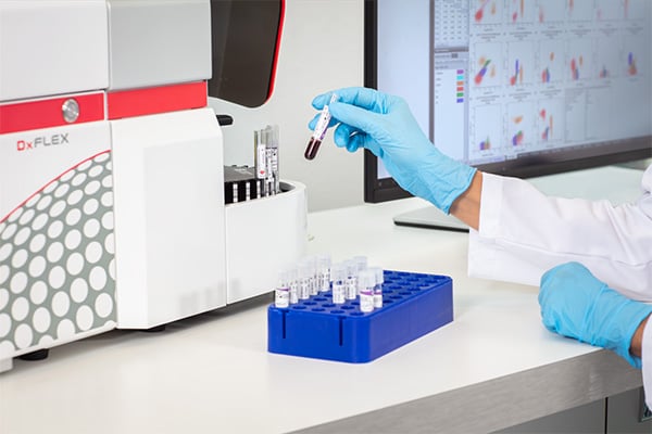
Analyze millions of cells per run
The DxFLEX flow cytometer helps you to analyze large cell populations with up to 25M events/file and 30K events/sec.
- Find the "needle in the haystack."
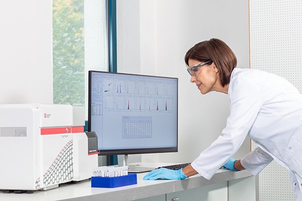
Paperless data transmission
Bi-directional LIS connectivity for transferring Specimen ID and test request from the LIS to the DxFLEX instrument, and sending results back to the LIS.
- Full connectivity for the digital world.
Introducing the DxFLEX Flow Cytometer
Learn more about the DxFLEX System


Streamline Leukemia and Lymphoma* Analysis
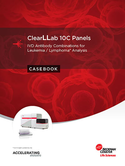
* For Non-Hodgkin’s lymphoma only
Explore DxFLEX Models
Technical Documents
Related Parts for DxFLEX
B73613, DxFLEX Sheath Fluid, 10 L
A nonionic, non-fluorescent, and azide-free sheath fluid for use on DxFLEX flow cytometers.

DxFLEX Autoloader Module
The optional autoloader module can be added to the DxFLEX flow cytometer to decrease hands on time during acquisition. The module accepts a standard 32-tube carousel, or with an additional optional adapter can support sample loading from 96-well plates*. When using the carousel, an integrated barcode reader can be used for extra verification of sample identity.
*Plate adapter: Regulatory status differs by region. Please contact your local sales representative.
DxFLEX Upgrade from B5-R0-V0 to B5-R3-V5
Activation key to upgrade the DxFLEX B5-R0-V0 flow cytometer with additional parameters. Upon installation the instrument will have 3 active lasers (488 nm, 638 nm, 405 nm) and 13 channels for fluorescence detection in the B5-R3-V5 configuration.
